Torn Meniscus Injury Treatment Knee Meniscus Repair Surgery
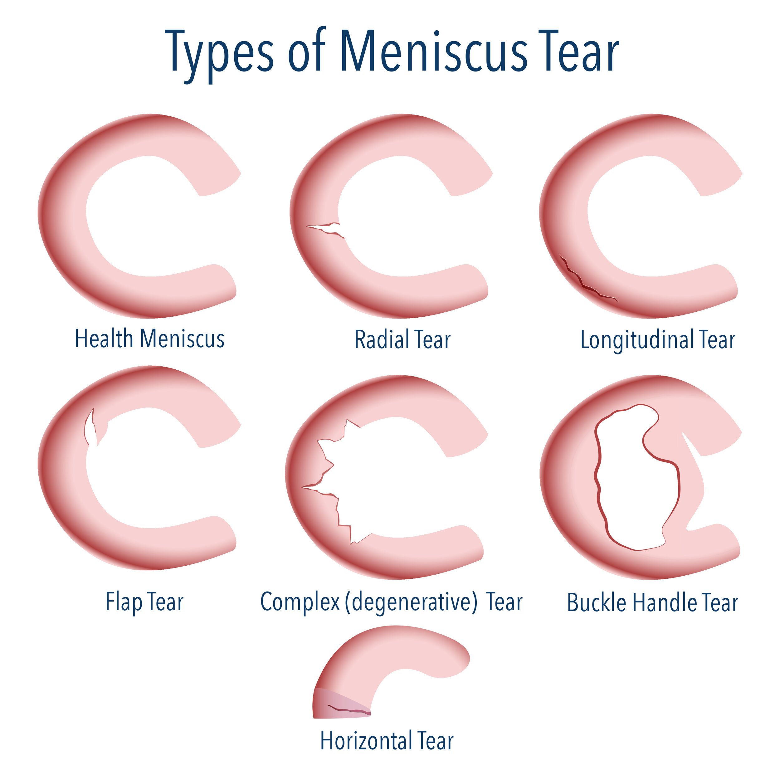
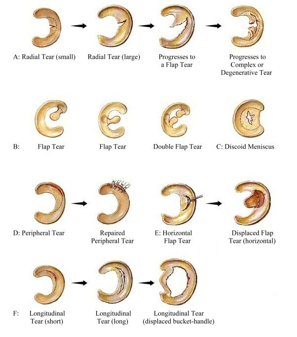
The meniscus a tissue that sits between the femur and tibia bone. It can tear in many different ways, and no two tears ever look the same. There are a few varieties frequently seen in MRI reports. Radial meniscus tear A radial tear is a tear across the fibers of the meniscus.

Illustrations of the meniscal root tear classification system in 5... Download Scientific Diagram
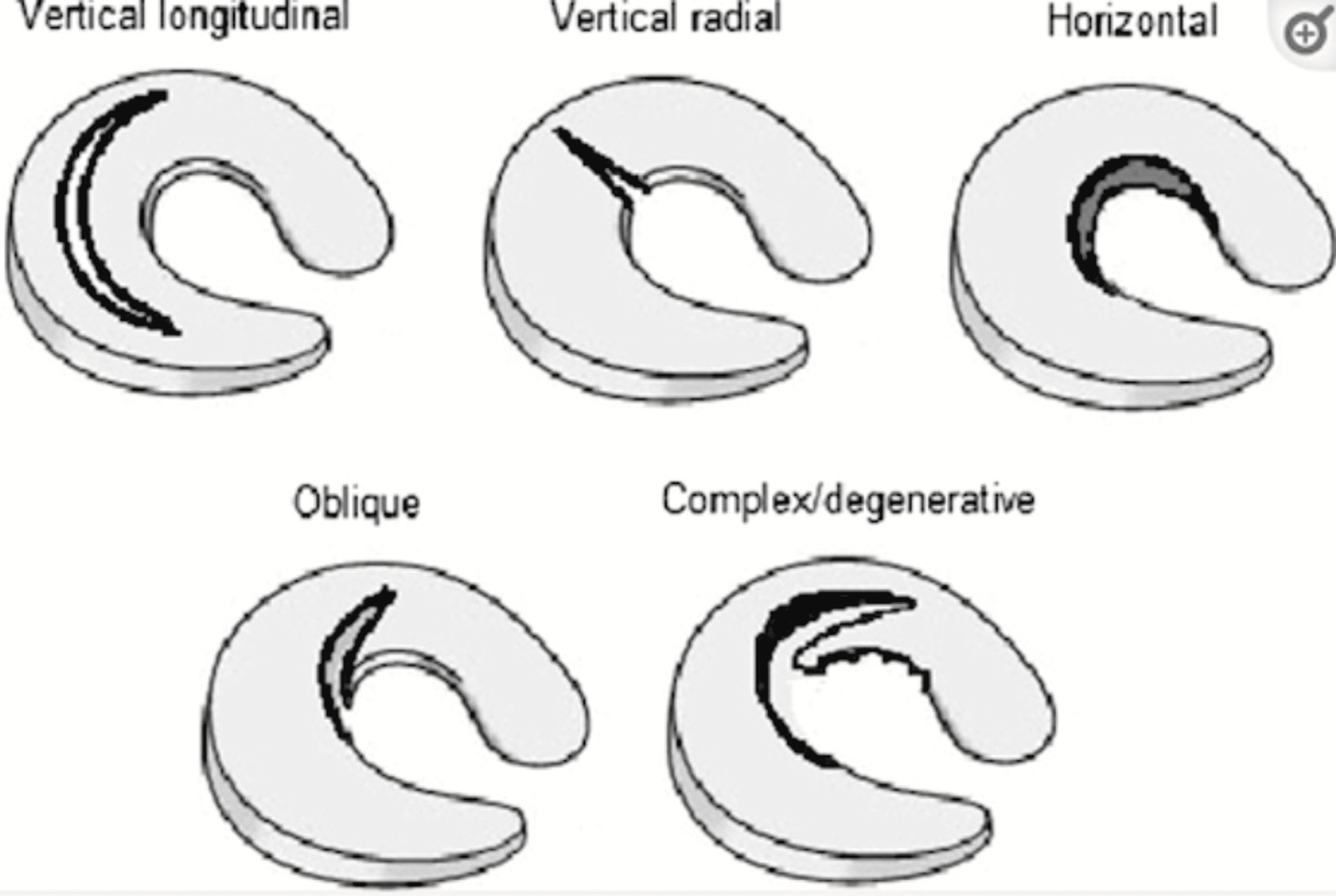
Meniscal ramp lesions consist in longitudinal vertical and/or oblique peripheral tears affecting the posterior horn of medial meniscus that may lead to meniscocapsular or meniscotibial disruption, in the setting of an ACL tear [].The coexistence of an ACL tear and other capsular and ligament injuries has been extensively described [].Acute ACL tear is associated with meniscal injuries in more.
/GettyImages-137278351-569c02f95f9b58eba4a700f8.jpg)
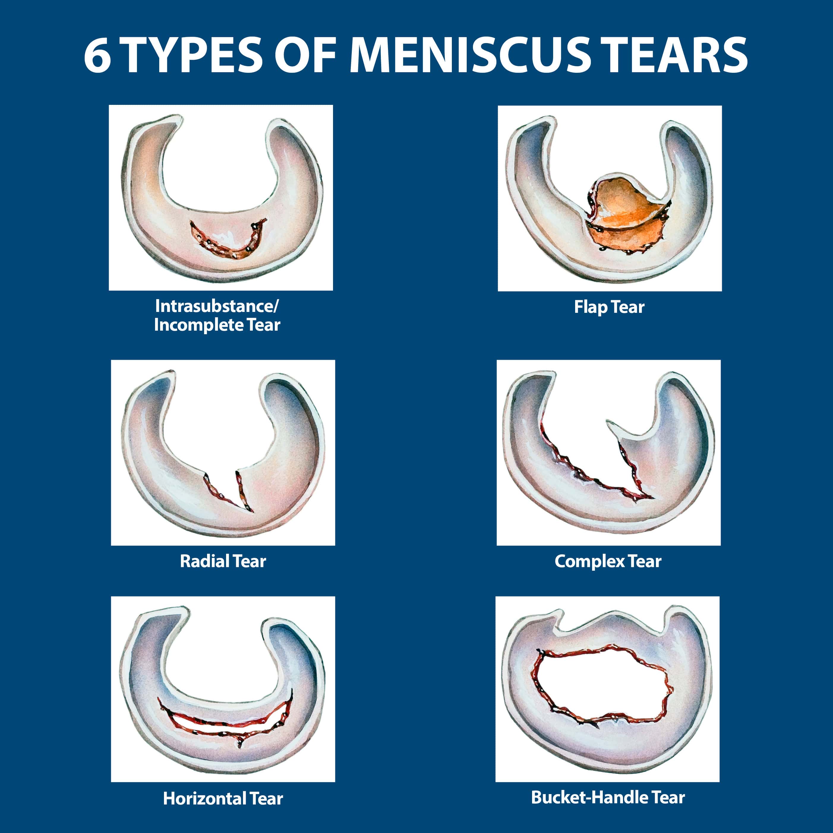
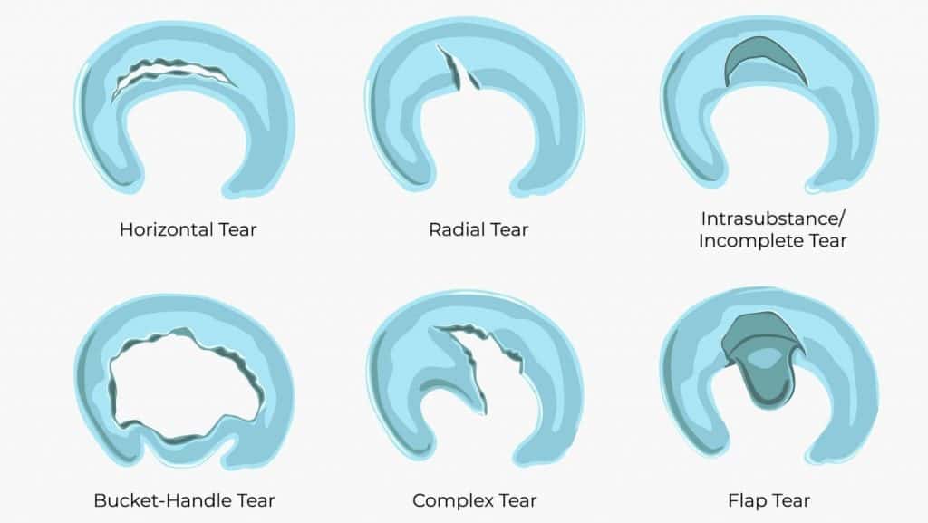
6 Types of Meniscus Tears and Locations
There are six types of meniscus tears: radial, intrasubstance, horizontal, flap, complex, and bucket-handle. All can compromise the knee, where this C-shaped cartilage is found. The part of the meniscus these tears affect, the patterns they exhibit, and their complexity differ, however.

Meniscal Tear Causes, Presentation and Treatment Bone and Spine
Though initially described as a functionless remain of a leg muscle [ 1 ], extensive scientific investigations in recent decades have described the meniscus as a vital part of the knee joint with anatomical, biomechanical, and functional importance [ 2 ].

Types of Meniscal Tears
Meniscal tears are common sports-related injuries in young athletes and can also present as a degenerative condition in older patients. Diagnosis can be suspected clinically with joint line tenderness and a positive Mcmurray's test, and can be confirmed with MRI studies.
Cureus Meniscus Tear Pathology, Incidence, and Management
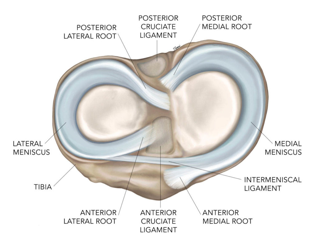
The menisci — the medial meniscus and lateral meniscus - are crescent-shaped bands of thick, rubbery cartilage attached to the shinbone (tibia). They act as shock absorbers and stabilize the knee. The medial meniscus is on the inner side of the knee joint. The lateral meniscus is on the outside of the knee.
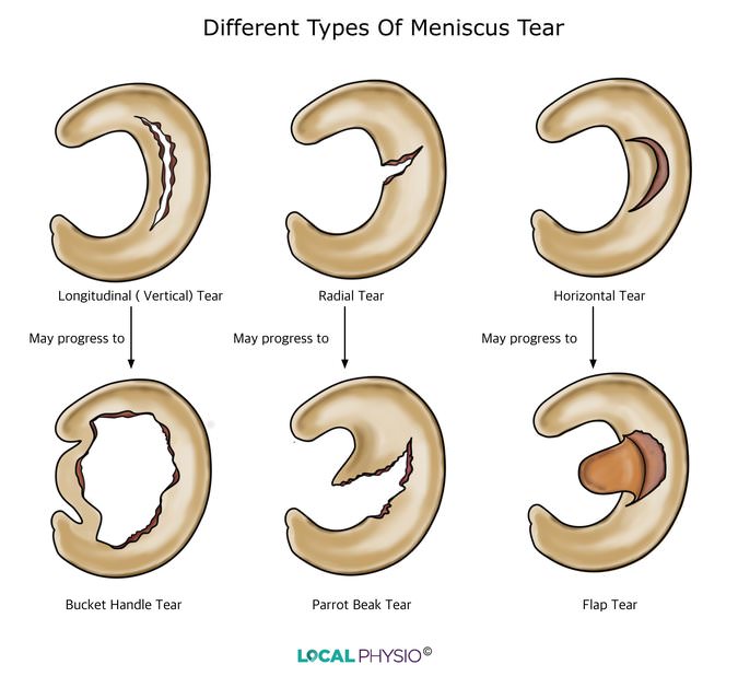
Meniscus Tear Local Physio
Occasionally, meniscal tears can be difficult to detect at imaging; however, secondary indirect signs, such as a parameniscal cyst, meniscal extrusion, or linear subchondral bone marrow edema, should increase the radiologist's suspicion for an underlying tear. Awareness of common diagnostic errors can ensure accurate diagnosis of meniscal tears.

MR Imagingbased Diagnosis and Classification of Meniscal Tears RadioGraphics
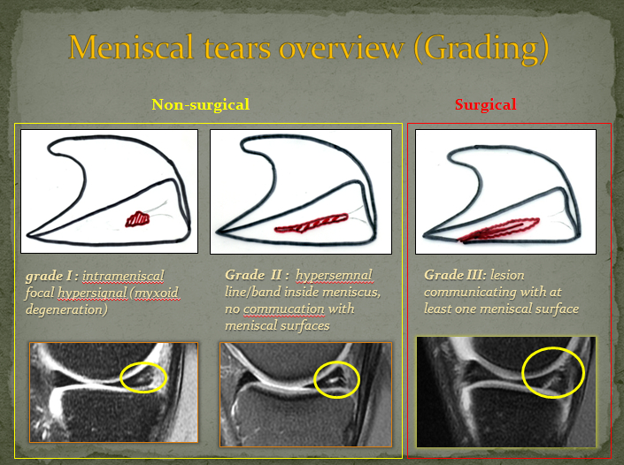
. MRI grading system classifies tears based on their appearance on an MRI scan (Fig. 8). Grade 0 represents an intact, normal meniscus. Grade I and Grade II signals do not intersect.
Meniscal Tears Brisbane Knee and Shoulder Clinic Dr MacgroartyBrisbane Knee and Shoulder Clinic
Meniscal injuries are a common problem in sports; they are the most frequent injury to the knee joint. Such injuries are especially prevalent among competitive athletes, particularly those.
Meniscal Root Tears Dr. Chris Jones Colorado Springs, CO
Meniscus tears are a common orthopedic pathology and planning a single, effective treatment is challenging. The diagnosis of meniscal tears requires detailed history-taking, physical examinations, special diagnostic tests, and most likely magnetic resonance imaging (MRI) to confirm the lesion.
EPOS™
Classification Grade 1 to 3 have been described on MRI: grade 1: small focal area of hyperintensity, no extension to the articular surface grade 2: linear areas of hyperintensity, no extension to the articular surface 2a: linear abnormal hyperintensity with no extension to the articular surface
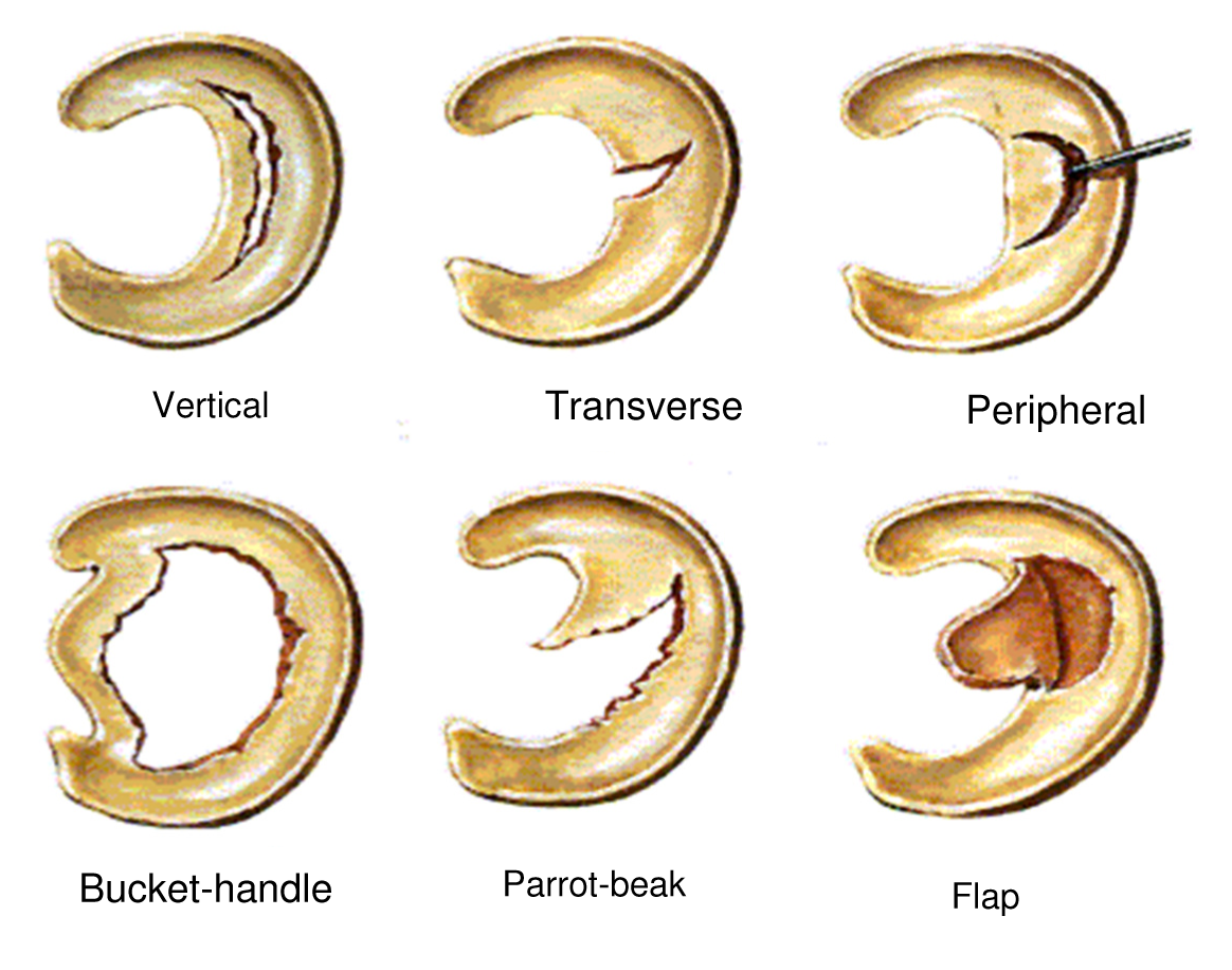
Meniscal Tear Types
Normal meniscus has uniformly low signal intensity on T2-weighted images (T2W). Grade I and II lesions can be a normal appearance of ageing in older patients. Classifications, online calculators, and tables in radiology. Martin C, Crues JV 3rd, Kaplan L, Mink JH. Meniscal tears: pathologic correlation with MR imaging. Radiology. 1987 Jun;163.

MR Imagingbased Diagnosis and Classification of Meniscal Tears RadioGraphics
Symptoms & causes Diagnosis & treatment Doctors & departments On this page Diagnosis Treatment Self care Preparing for your appointment Diagnosis A torn meniscus often can be identified during a physical exam.
Lateral Meniscus Tear Symptoms, Causes and Diagnosis
Meniscal tears are a common pathology and diagnosis relies on a detailed clinical history and clinical examination, magnetic resonance imaging (MRI), and arthroscopy. Some types of meniscal tears (e.g. horizontal or oblique tears) may not always be related to clinical symptoms, and they are frequently encountered in asymptomatic knees [ 1 ].
Mediale meniscus Creative Saplings
A meniscus root tear may occur in circumstances that appear benign, such as walking up a flight of stairs or stepping off a curb. It also can occur with a sports activity such as pivoting in pickleball or lifting a heavy kayak into the water. Athletes also incur meniscus root tears with an accompanying ACL tear in a more traumatic injury.

Grade 1 Meniscal signal or tear( MRI finding) and Grade 2 Meniscal Tear YouTube
Meniscal tears are the failure of the fibrocartilaginous menisci of the knee. There are several types and can occur in an acute or chronic setting. Meniscal tears are best evaluated with MRI. Pathology
